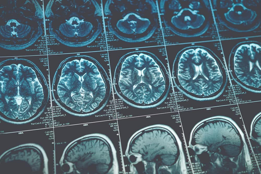
At Ambrosia in West Palm Beach, FL, we are committed to exploring cutting-edge methods for understanding and improving mental health. One such groundbreaking technique is brain mapping, a revolutionary process that allows us to visualize the brain’s activity in intricate detail. By identifying patterns and anomalies in brain function, brain mapping provides invaluable insights into an individual’s mental health and behavioral tendencies.
This innovative approach not only enhances our understanding of complex mental health issues but also guides us in designing more personalized and effective treatment plans. Join us as we delve into the fascinating world of brain mapping and discover how it can help unlock a healthier, more balanced life.
Understanding the Concept of Brain Mapping

The Science Behind Brain Mapping
The foundational science of brain mapping lies in utilizing neuroimaging technologies such as Magnetic Resonance Imaging (MRI), Electroencephalography (EEG), and Positron Emission Tomography (PET). These technologies provide valuable data on brain activity and structure, offering insights into how neural pathways engage during different tasks or in response to stimuli.
Furthermore, advances in computational models and artificial intelligence have enhanced our ability to analyze and visualize this data, revealing intricate patterns of connectivity that may influence behavior, emotion, and cognition. For instance, machine learning algorithms can now sift through vast amounts of neuroimaging data to identify biomarkers associated with conditions such as Alzheimer’s disease or depressive disorders, paving the way for earlier diagnosis and more personalized treatment plans.
The Evolution of Brain Mapping Techniques
Historically, brain mapping has evolved from rudimentary approaches such as lesion studies and simple observational methods to advanced imaging techniques that can visualize brain activity in real-time. The launch of fMRI in the early 1990s marked a significant milestone, enabling non-invasive monitoring of brain activity while subjects performed tasks or exhibited thoughts.
Today, methods like diffusion tensor imaging (DTI) and magnetoencephalography (MEG) are utilized in research and clinical practices, leading to a better understanding of both healthy and disease-affected brains. These technologies not only allow for the mapping of brain structure but also provide insights into the timing of neural activity, which is crucial for understanding processes such as decision-making and sensory perception. As these techniques continue to advance, they hold the promise of unlocking even deeper insights into the complexities of the human brain, potentially revolutionizing fields such as psychology, psychiatry, and neurology.
The Importance of Brain Mapping
The significance of brain mapping extends across various fields, particularly in medicine and psychology. By revealing the underlying mechanisms of brain function, researchers can tailor interventions and assess outcomes effectively. This knowledge bolsters the development of new treatments and enhances diagnostic practices, making brain mapping a crucial aspect of modern neuroscience.
In medical science, brain mapping plays a pivotal role in diagnosing and treating neurological disorders such as epilepsy, Alzheimer’s disease, and multiple sclerosis. By mapping specific brain areas associated with these conditions, healthcare professionals can identify abnormalities early and formulate personalized treatment plans.
It also informs surgical approaches—such as pre-surgical brain mapping—where understanding brain function is vital to avoiding damage during procedures treating tumors or epilepsy sites. Techniques like functional MRI (fMRI) and electrocorticography allow surgeons to visualize brain activity in real-time, ensuring that critical areas responsible for speech, movement, and other functions are preserved during operations. This precision not only increases the success rates of surgeries but also significantly enhances the quality of life for patients post-operation.
Brain mapping is equally essential in psychological research, helping psychologists explore complex phenomena such as cognition, emotion, and behavior. Through various studies, researchers utilize brain mapping to investigate how different brain regions influence mental health, contributing to evidence-based practices in therapy and counseling.
This growing field of research aids in identifying biomarkers for psychological disorders, facilitating more effective diagnosis and intervention protocols. For instance, neuroimaging studies have shown distinct patterns of brain activity associated with conditions like depression and anxiety, leading to targeted therapeutic approaches such as cognitive-behavioral therapy or pharmacological treatments tailored to individual brain profiles. The insights gained from brain mapping not only enhance our understanding of mental health disorders but also pave the way for innovative strategies in prevention and treatment, ultimately aiming to improve patient outcomes and well-being.
Brain mapping plays a pivotal role in behavioral health treatment by providing a detailed visualization of brain activity, which is essential for understanding the complexities of mental health conditions. By identifying specific patterns and anomalies in the brain’s function, this advanced technology enables healthcare professionals to gain deeper insights into the root causes of various mental health issues. Such information is invaluable in diagnosing conditions more accurately and crafting personalized treatment plans that cater to the unique needs of each patient.
This tailored approach not only improves patient outcomes but also enhances the effectiveness of different types of therapies. Moreover, brain mapping holds the potential to unlock new perspectives on behavioral health, leading to more targeted and innovative interventions. By integrating brain mapping into treatment strategies, practitioners can offer more precise and effective care, paving the way for a brighter future in mental health management.
What is the Process of Brain Mapping?
The process of brain mapping involves several steps to ensure accurate visualizations and analyses of brain activity and structure. From selecting the appropriate tools and techniques to interpreting data, each phase contributes to a comprehensive understanding of the brain.
Tools and Techniques Used
There are a variety of tools and techniques employed in brain mapping, including but not limited to:
- Functional Magnetic Resonance Imaging (fMRI)
- Electroencephalography (EEG)
- Magnetoencephalography (MEG)
- Positron Emission Tomography (PET)
- Diffusion Tensor Imaging (DTI)
- Computerized Tomography (CT) scans
Each method has its distinct advantages and limitations, making the selection of the appropriate technique essential based on the research question and desired outcomes. For instance, while fMRI is excellent for mapping brain activity related to specific tasks due to its high spatial resolution, EEG offers superior temporal resolution, making it ideal for capturing rapid changes in brain activity. This interplay between spatial and temporal resolution is crucial for researchers aiming to understand the dynamics of neural processes.
Steps Involved in Brain Mapping
The steps involved in brain mapping typically include:
- Identifying the research question or clinical need.
- Selecting the appropriate neuroimaging technique.
- Preparing the participants and ensuring ethical compliance.
- Conducting the imaging procedure.
- Analyzing the collected data using advanced statistical methods.
- Interpreting the results in the context of the original research question.
This methodical approach ensures the validity and reliability of the findings, enhancing the overall quality of the research outcomes. Additionally, the preparation of participants is a critical phase that often involves obtaining informed consent, screening for contraindications, and providing detailed instructions to minimize anxiety and ensure cooperation during the imaging process. Furthermore, the analysis phase can incorporate sophisticated machine learning algorithms to detect patterns in the data that may not be immediately apparent, thus enriching the insights gained from the study.
What are the Different Types of Brain Mapping?

Functional Brain Mapping
Functional brain mapping measures brain activity associated with specific tasks or stimuli. Techniques such as fMRI and EEG are widely used to visualize active brain areas in real time, showcasing how various regions coordinate during cognitive processes.
This type of mapping is particularly useful for understanding brain networks involved in memory, emotion regulation, and decision-making, enabling researchers to investigate the intricacies of human behavior. For instance, fMRI has been instrumental in uncovering how different brain areas communicate during complex tasks, such as language processing or problem-solving, revealing the dynamic interplay between regions that were once thought to operate independently. Furthermore, advancements in machine learning algorithms are beginning to allow for the prediction of cognitive states based on brain activity patterns, opening new avenues for understanding mental health conditions and cognitive impairments.
Structural Brain Mapping
In contrast, structural brain mapping involves the examination of the anatomy of the brain, including its physical structures and pathways. Techniques like MRI allow for detailed imaging of brain regions, assisting in identifying abnormalities in structure that may correlate with neurological and psychiatric disorders.
Understanding brain structure is fundamental in both research and clinical settings, providing information essential for both diagnosis and treatment planning. For example, diffusion tensor imaging (DTI), a specific type of MRI, maps the white matter tracts connecting different brain regions, offering insights into how these connections may be disrupted in conditions such as multiple sclerosis or traumatic brain injury.
Additionally, structural mapping has paved the way for studies on neuroplasticity, illustrating how the brain can reorganize itself in response to learning, injury, or environmental changes, thereby enhancing our understanding of recovery processes in various neurological conditions.
Challenges and Limitations of Brain Mapping
While brain mapping presents numerous advantages, it also faces challenges and limitations that researchers and clinicians must consider. Addressing these issues is essential for advancing the field and ensuring ethical practices in neuroscience.
Ethical Considerations
Brain mapping raises several ethical concerns, particularly regarding the privacy and consent of participants. Researchers must navigate the delicate balance between acquiring necessary data and respecting individual autonomy and confidentiality. The implications of findings from brain mapping studies can also impact perceptions of mental illness, cognition, and identity.
Ethical frameworks and guidelines are crucial for ensuring that research is conducted responsibly fostering public trust in scientific inquiry. Furthermore, the potential misuse of brain mapping technologies poses a significant risk. For instance, the ability to interpret brain activity could lead to invasive practices or discrimination based on neurological data. This necessitates a robust dialogue among ethicists, neuroscientists, and policymakers to establish clear regulations that protect individuals while promoting scientific progress.
Technological Constraints
Additionally, technological constraints pose challenges in brain mapping. While advancements have significantly improved the resolution and accuracy of imaging techniques, limitations still exist. Issues such as artifacts in imaging, varying interpretations of results, and the high cost of sophisticated equipment can hinder the accessibility and applicability of brain mapping techniques.
Overcoming these constraints requires ongoing investment in research, innovation, and collaboration within the neuroscience community to enhance methods and tools for brain mapping. Moreover, there is a growing need for interdisciplinary approaches that integrate insights from fields like artificial intelligence and machine learning to improve data analysis and interpretation.
Ambrosia Can Help You Recover
As we wrap up our exploration into the transformative world of brain mapping, it’s clear that this innovative technique is reshaping our understanding of mental health. By illuminating the intricate workings of the brain, brain mapping provides crucial insights that aid in crafting personalized treatment programs tailored to each individual’s unique needs.
At Ambrosia in South Florida, we are proud to be at the forefront of applying these advanced methodologies to enhance mental well-being. Whether you’re seeking answers for yourself or a loved one, our dedicated team is here to guide you through this enlightening journey. Don’t hesitate to reach out to us for more information or to schedule a consultation and take the first step towards a brighter, healthier future.


